


Diseases
- Allergic Conjunctivitis
- Behcet Disease
- Blepharoshalasis Dermatochalases
- Diabetic Retinopathy
- Ectropion (Eversion of the Eyelids)
- Entropion (Inversion of the Eyelids)
- Epiretinal Membrane
- Episcleritis
- Glaucoma
- The Anatomy Of The Eye
- Intraocular Bleddings
- Eyelid Inflammations
- Xerophthalmia
- Injuries In The Eye
- Lachrymal Duct Obstruction
- Floaters
- Herpetic Ceratitis
- Cataract
- Keratoconus
- Refraction
- Macular Hole
- Macular Edema
- Microbial Keratitis
- Microbial Conjunctivitis
- Optic Neuritis and Multiple Sclerosis
- Presbyopia
- Pterygium
- Ptosis (Looseness Of The Eyelid)
- Color Blindness
- Retinal Detachment
- Retinal Embolism
- Retinitis Pigmentosa
- Retinoblastoma
- Yellow Spot Disease (ARMD)
- Scleritis
- Chalazion
- Thyroid Orbitopathy
- Uveitis
- Keratopathy Caused By Bells Palsy
Retinoblastoma
DEFINITION: Retinoblastoma is a malignant tumor formed in the nerve layer of the retina. It is the most common intraocular tumor of childhood with a prevalence of 1 out of 17000. Loss of vision is inevitable if the patient is late for treatment, and unfortunately, some cases result in deaths. Most patients have a family history. Patients are generally diagnosed at the age of 1 or 2 years, although some cases can be detected later.
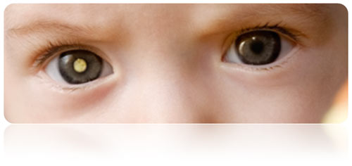
Figure 1. Leukocoria in an infant.
RISK FACTORS AND CAUSES: 40% of retinoblastomas are genetic. In 1 out of 3-4 cases, it is observed that both eyes are involved. Involvment of both eyes can be at the same time or develop later. The healthy eye should be checked frequently. Siblings of the patient should be examined for this disease.
FINDINGS: The most common clinical finding of retinoblastoma is the pupil losing its normal red reflection and shining white (leukocoria). If a whiteness is seen in the child's pupil by the naked eye or in a photograph taken by flashlight, tumor should be suspected and an ophthalmologist should be consulted immediately. Another common finding is strabismus. Sometimes, the tumor may push the eye outward.
Figure 2. Fundus photography of retinoblastoma (left). Posterior segment (middle) and optical coherence tomography imaging (right) of a patient diagnosed with spontaneously regressed retinoblastoma.
